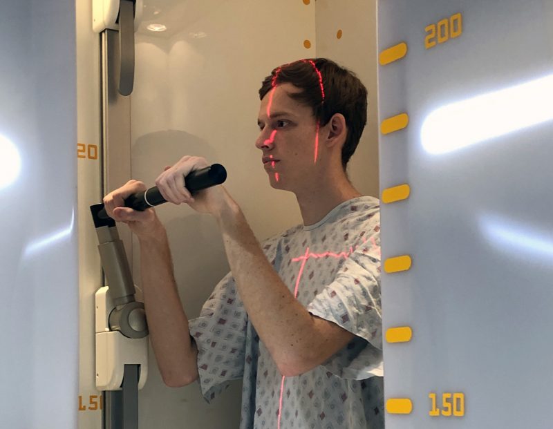
While being treated for scoliosis, Bailey Fumich’s exposure to radiation will be significantly reduced thanks to new technology in our Orthopedics center.
Bailey Fumich’s mother has lost count of how many X-rays he’s had to have since first being seen for scoliosis nearly a decade ago.
“He’s had a lot,” said Tina McKenna, noting that Bailey got X-rays about every 6 months as doctors in the Akron Children’s spine program monitored his scoliosis.
Now Bailey, who underwent spinal fusion surgery in early December, is benefiting from the hospital’s new EOS™ machine, which captures X-ray images using significantly lower doses of radiation. Akron Children’s is the first hospital in Northeast Ohio to install the technology.
Bailey, 17, and his parents agreed that him being exposed to less radiation as he continues his follow up at the hospital is a good thing. According to Dr. Lorena Floccari, one of the orthopedic surgeons who performed his surgery, Bailey will need X-rays at his 3- and 6-month visits and at 1-, 2- and 5-year visits post-surgery.

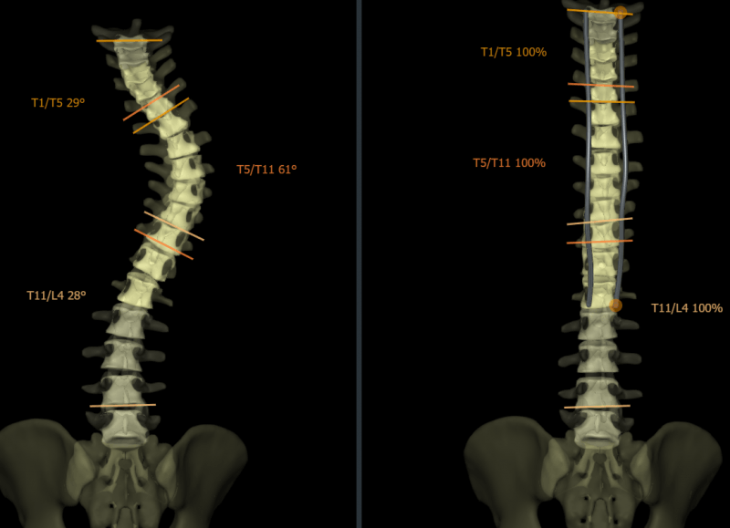
Side-by-side, 3D EOS images show Bailey’s spine after and before scoliosis surgery.
Orthopedics is using the EOS for patients with spine disorders like scoliosis. The EOS can also be used to get 2D and 3D images of the whole leg or shoulders, such as to evaluate a leg length discrepancy or limb deformity.
According to the company, the EOS system offers 50% to 85% less radiation than traditional X-ray systems and 95% less dose than basic computed tomography (CT) scans, in accordance with the ALARA (As Low As Reasonably Achievable) principle for minimizing a patient’s exposure to radiation.
Reducing radiation dose is particularly beneficial for children requiring frequent imaging, like Bailey. The Micro Dose feature further reduces radiation exposure, offering frontal and lateral pediatric full spine images at a dose that’s equivalent to only a week’s worth of natural radiation.
EOS captures bi-planar images with 2 perpendicular X-ray beams that travel vertically while scanning the patient from head to toe. In less than 20 seconds, the EOS exam produces simultaneous frontal and lateral low-dose images. The 2 resulting digital images are processed by EOS’ proprietary sterEOS® software to generate a 3D model of the patient’s spine and/or lower limbs. These 3D models provide highly detailed information about the patient’s unique anatomy to better assist orthopedic surgeons as they diagnose patients. This additional data can be used in the EOSapps for precise, 3D surgical planning to help improve overall patient outcomes by identifying patients at risk and developing a customized surgical plan.
The system is so safe that parents can be with their children in the room while it’s being used, unlike with a traditional X-ray.
 Dr. Floccari used EOS previously during her fellowship in spinal surgery in New Zealand, so she was happy to see that Akron Children’s has added the technology.
“This improves our diagnostic capabilities while providing the highest level of safety to our patients,” she said. “We are thrilled to offer this new technology to patients with a wide range of spine and other orthopedic conditions.”
This service is currently offered on our Akron campus. Learn more about EOS imaging and the treatments offered at the Spine Center , an Akron Children’s Center of Excellence.


